Author: Justine Ko, MD
Peer-reviewer: Terese Whipple, MD
Final editor: Alex Tomesch, MD
A 32-year old female presents to the emergency department with right ankle pain after a high speed motor vehicle accident. On exam, she is noted to have ecchymosis and swelling over the dorsal foot, and pain with ankle dorsiflexion and plantarflexion.

Image 1. Case courtesy of Dr Charlie Chia-Tsong Hsu, Radiopaedia.org, rID: 18235
What is your diagnosis?
What is your management in the ED?
What is your disposition? Do you consult orthopaedics emergently?
-- see below for answers --
What is your diagnosis?
The patient is diagnosed with a talar neck fracture. These fractures are associated with high-impact mechanisms and account for nearly 50% of all talar fractures.[1] X-ray is a good initial imaging modality for these injuries, however CT should be considered to better assess the articulating surfaces of the talus.
Pearl: To aid in viewing the talar neck, a Canale view can be used. The x-ray is taken with the ankle plantar flexed and pronated 15˚. The beam of the x-ray should be angled 75˚ from the horizontal plane where the foot lies.[2]
What is your management in the ED?
Management depends on the type of talar neck fracture. The most common classification system for talar neck fractures is the Hawkins-Canale classification:
Type I fractures: nondisplaced fractures that usually only require splinting and non-weight bearing status.
Type II-IV fractures are displaced fractures with an associated dislocation that requires urgent reduction in the emergency department
Type II fractures: talocalcaneal dislocation
Type III fractures (Image 2): talocalcaneal and talotibial dislocations
Type IV: all talar articulations disrupted.[3,4]
Pearl: Most talar injuries are usually caused by forced hyperdorsiflexion of the ankle. They are often associated with other injuries in the patients and frequently present as an open fracture.[1] A detailed secondary survey and skin exam should be performed on all patients.
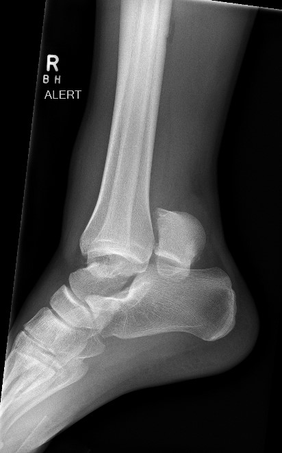
Image 2: Case courtesy of Assoc Prof Craig Hacking, Radiopaedia.org, rID: 84976
3. What is your disposition? Do you consult orthopaedics emergently?
For Type I fractures, close orthopedic followup can be arranged after the patient is splinted in a posterior mold splint. Given the association with high-impact mechanisms, type I fractures are rare. Type II-IV fractures require orthopedic consultation and urgent reduction. These fractures are associated with increased risk for osteonecrosis given the disruption of the talar blood supply with dislocation.[1]
References:
Shamrock AG, Byerly DW. Talar Neck Fractures. In: StatPearls. StatPearls Publishing; 2021. Accessed June 19, 2021. http://www.ncbi.nlm.nih.gov/books/NBK542315/1.
Melenevsky Y, Mackey RA, Abrahams RB, Thomson NB. Talar Fractures and Dislocations: A Radiologist's Guide to Timely Diagnosis and Classification. Radiographics. 2015;35(3):765-779. doi:10.1148/rg.2015140156
Dale JD, Ha AS, Chew FS. Update on Talar Fracture Patterns: A Large Level I Trauma Center Study. American Journal of Roentgenology. 2013;201(5):1087-1092. doi:10.2214/AJR.12.9918
Alton T, Patton DJ, Gee AO. Classifications in Brief: The Hawkins Classification for Talus Fractures. Clin Orthop Relat Res. 2015;473(9):3046-3049. doi:10.1007/s11999-015-4136-x

Sign up here to receive our weekly cases
Want to write? Email us at orthoempearls@gmail.com
Instagram/Twitter: @orthoempearls
Comments
Post a Comment